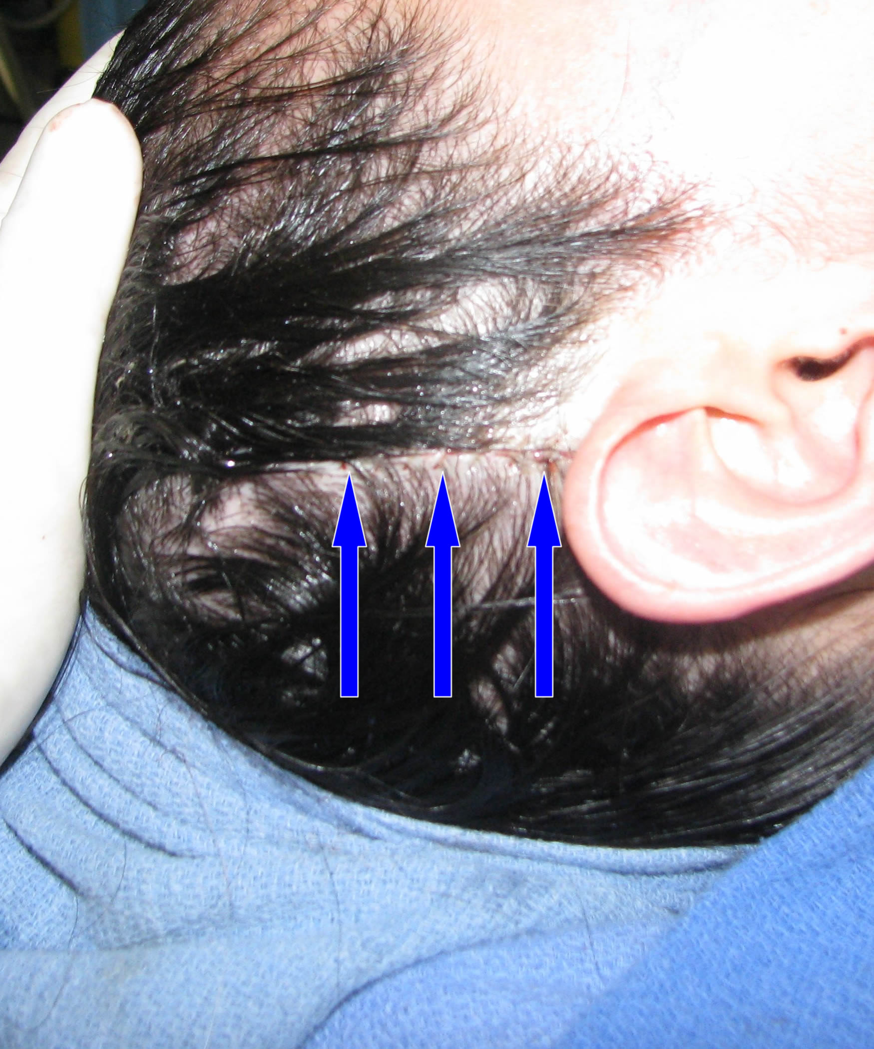
I happen to perform fascia grafting as part of rhinoplasty and revision rhinoplasty on a fairly regular basis. I do this type of grafting mostly in rhinoplasty patients with thin skin. In this patient population, anything done to reshape the nose can cause unwanted visible contour irregularities post-operatively because thin skin has a tendency to rapidly shrink wrap down to the new shape. In order to minimize risks of visible contour irregularities after rhinoplasty or revision rhinoplasty, I place a layer of fascia under the skin and over the cartilage or bone. This helps to bolster the thickness of the skin and reduces chances that even minor contour irregularities can be seen as the skin shrink wraps down. The fascia is essentially a thin layer or sheet of connective tissue that can be found in many areas of the body. I happen to use temporal fascia for my grafting since it is readily available and in plentiful supply. Many patients inquire about the incision used for the harvesting of the temporal fascia in rhinoplasty. I provided the photo below to visually demonstrate to potential patients where the actual incision is made. I performed a rhinoplasty with temporal fascia grafting on this patient just yesterday, which prompted me to post a quick blog entry. As you can see, I make a vertical incision above the right ear, which is completely contained within the hair (see blue arrows). Once this heals, it is incredibly difficult to even see it. I don’t shave any hair to make the incision, which is a common concern among patients getting ready to have this done. The photo below shows the actual incision after I have closed it with absorbable sutures (meaning no sutures have to be removed). The incision tends to heal quite nicely and after a week or so most rhinoplasty patients don’t even really remember having had the fascia grafting portion of the surgery.

